Knee
- Anatomy
- Conditions
- Procedures
Knee Anatomy
The knee is a complex joint made up of different structures including bones, tendons, ligaments and muscles. They all work together to maintain normal function and provide stability to the knee during movement.
Having a well-functioning healthy knee is essential for our mobility and ability to participate in various activities. Understanding the anatomy of the knee enhances your ability to discuss and choose the right treatment procedure for knee problems with your doctor.
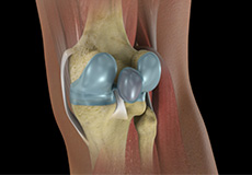
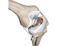
Bones of the Knee
The knee is a hinge joint made up of two bones, the thighbone (femur) and the shinbone (tibia). There are two round knobs at the end of the femur called femoral condyles which articulate with the flat surface of the tibia called the tibial plateau. The tibia plateau on the inside of the leg is called the medial tibial plateau, and on the outside of the leg it is called the lateral tibial plateau.
The two femoral condyles form a groove on the front (anterior) side of the knee called the patellofemoral groove. A small bone called the patella sits in this groove and forms the knee cap. It acts as a shield and protects the knee joint from direct trauma.
A fourth bone called the fibula is the other bone of the lower leg. This form a small joint with the tibia. This joint has very little movement and is not considered a part of the main joint of the knee.
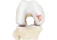
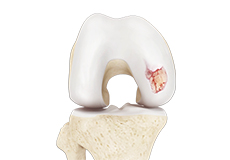
Articular Cartilage and Menisci of the Knee
Movement of the bones causes friction between the articulating surfaces. To reduce this friction, all articulating surfaces involved in movement are covered with a white, shiny, slippery layer called articular cartilage. The articulating surface of the femoral condyles, tibial plateaus and the back of the patella are covered with this cartilage. The cartilage provides a smooth surface that facilitates easy movement.
To further reduce friction between the articulating surfaces of the bones, the knee joint is lined by a synovial membrane which produces a thick clear fluid called synovial fluid. This fluid lubricates and nourishes the cartilage and bones inside the joint capsule.
Within the knee joint between the femur and tibia there are two C shaped cartilaginous structures called menisci. Menisci function to provide stability to the knee by spreading the weight of the upper body across the whole surface of the tibial plateau. The menisci help in load bearing by preventing the weight from concentrating onto a small area, which could damage the articular cartilage. The menisci also act as a cushion between the femur and tibia by absorbing the shock produced by activities such as walking, running and jumping.
Ligaments of the Knee
Ligaments are tough bands of tissue that connect one bone to another bone. The ligaments of the knee function to stabilize the knee joint. There are two important groups of ligaments that hold the bones of the knee joint together, collateral ligaments and the cruciate ligament.
Collateral ligaments are present on either side of the knee. They function to prevent the knee from moving too far during side to side motion. The collateral ligament on the inside is called the medial collateral ligament (MCL) and the collateral ligament on the outside is called the lateral collateral ligament (LCL).
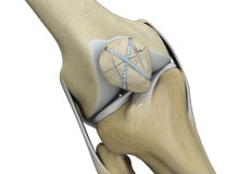
Cruciate ligaments present inside the knee joint, control the back and forth motion of the knee. The Cruciate ligament in the front of the knee is called anterior cruciate ligament or ACL and the cruciate ligament in the back of the knee is called posterior cruciate ligament or PCL.
Muscles of the Knee
There are two major muscles, the quadriceps and the hamstrings, which enable movement of the knee joint. The quadriceps muscles are in the front of the thigh. When the quadriceps muscles contract, the knee straightens. The hamstrings are in the back of the thigh. When the hamstring muscles contract, the knee bends.
Tendons of the Knee
Tendons are structures that attach muscles to the bone. The quadriceps muscles of the knee meet just above the patella and attach to it through a tendon called the quadriceps tendon. The patella further attaches to the tibia through a tendon called the patella tendon. The quadriceps muscle, quadriceps tendon and patellar tendon all work together to straighten the knee. Similarly, the hamstring muscles at the back of the leg are attached to the knee joint with the hamstring tendon.
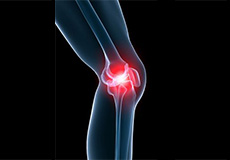
Knee Pain
The knee is one of the largest joints in the body, formed by the lower end of the femur, upper end of the tibia and the patella or knee cap. Several ligaments and muscles attach to the bones of the knee joint to maintain normal motion of the joint. Special cartilaginous tissues known as menisci are placed between the two articular ends of the joint. These act as a cushion between the articular surfaces and absorb the shock during movement.
Baker's Cyst
The knee consists of a fluid called synovial fluid, which reduces friction between the bones of the knee joint while you move your leg. Sometimes this fluid is produced in excess, resulting in its accumulation in the back of your knee. A Baker’s cyst or popliteal cyst is a fluid-filled swelling that develops into a lump behind the knee. This causes stiffness, tightness and pain behind your knee. It is commonly seen in women and people aged over 40 (although it can develop at any age).
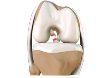
ACL Tears
The anterior cruciate ligament, or ACL, is one of the major ligaments of the knee that is in the middle of the knee and runs from the femur (thighbone) to the tibia (shinbone). It prevents the tibia from sliding out in front of the femur. Together with posterior cruciate ligament (PCL) it provides rotational stability to the knee.
MCL Tears
The medial collateral ligament (MCL) is the ligament that is located on the inner part of the knee joint. It runs from the femur (thighbone) to the top of the tibia (shinbone) and helps in stabilizing the knee.
Meniscal Injuries
The knee is one of the most complex and largest joint in the body, and is more susceptible to injury. Meniscal tears are one among the common injuries to the knee joint. It can occur at any age, but are more common in athletes playing contact sports.
Ligament Injuries
The knee is a complex joint which consists of bone, cartilage, ligaments and tendons that make joint movements easy and at the same time more susceptible to various kinds of injuries.
Knee problems may arise if any of these structures get injured by overuse or suddenly during sports activities. Pain, swelling, and stiffness are the common symptoms of any damage or injury to the knee.
Knee Arthritis
Arthritis is a general term covering numerous conditions where the joint surface or cartilage wears out. The joint surface is covered by a smooth articular surface that allows pain free movement in the joint. This surface can wear out for several reasons; often the definite cause is not known.
Osteoarthritis
Osteoarthritis, also called degenerative joint disease, is the most common form of arthritis. It occurs most often in older people. This disease affects the tissue covering the ends of bones in a joint (cartilage). In a person with osteoarthritis, the cartilage becomes damaged and worn out causing pain, swelling, stiffness and restricted movement in the affected joint.
Non-surgical Treatments

Physical Examination of the Knee
A complete physical examination of the knee is performed when you present to your doctor with a knee complaint. Both of your knees are examined and the results of the injured knee are compared to those of the healthy knee.
Surgical Treatment
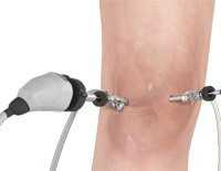
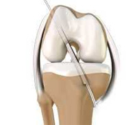
Knee Arthroscopy
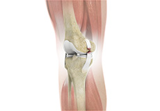
The knee joint is one of the most complex joints of the body. The lower end of the thighbone (femur) meets the upper end of the shinbone (tibia) at the knee joint. A small bone called the patella (kneecap) rests on a groove on the front side of the femoral end. A bone of the lower leg (fibula) forms a joint with the shinbone.
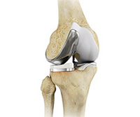
Minimally Invasive Knee Joint Replacement
Total knee replacement is a very successful surgical treatment for knee arthritis. Over the years, minimally invasive knee replacement surgical techniques have been developed to lessen tissue trauma and improve patient outcomes.
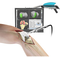
Computer Navigation for Total Knee Replacement
A total knee replacement surgery involves replacing the damaged surfaces of the articulating bones with the artificial implant. Most of these implants wear with use. Thus, the risk of need for revision surgery is high in young and active people if the implant must last the lifetime of the patient.
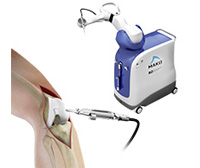
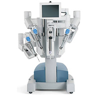
Mako Robotic-Arm Assisted Technology for Total Knee Replacement
We understand that making sure you know what to expect from your joint replacement experience is important to you. As you are reading through this material, if you have additional questions please reach out to us to discuss.
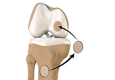
Unicompartmental Knee Replacement
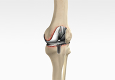
Unicompartmental knee replacement is a minimally invasive surgery in which only the damaged compartment of the knee is replaced with an implant. It is also called a partial knee replacement.
Robotic Assisted Partial Knee Surgery
Robotic Assisted Partial knee surgery is an innovative alternative to the conventional surgical procedure in patients suffering from degenerative knee diseases such as osteoarthritis. It is performed using robotic-arm technology that allows the surgeon to precisely perform the surgery through a smaller incision as compared to traditional surgery.
Revision Knee Replacement
Revision knee replacement surgery involves replacing part or all your previous knee prosthesis with a new prosthesis. Although total knee replacement surgery is successful, sometimes the procedure can fail due to various reasons and require a second revision surgery.
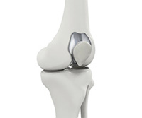
Patellofemoral Knee Replacement
Traditionally, a patient with only one compartment of knee arthritis would undergo a total knee replacement surgery. Patellofemoral knee replacement is a minimally invasive surgical option that preserves the knee parts not damaged by arthritis as well as the stabilising anterior and posterior cruciate ligaments, ACL and PCL.
Knee Reconstruction
The knee is the most complex joint in the body and is formed by the articulation between the thighbone (femur) and the shinbone (tibia). A kneecap is present over the front of the joint to provide extra protection. These bones are held together by four strong rope like structures called ligaments. Two collateral ligaments are present on either side of the knee and control the sideway movements of the knee.
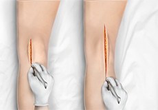
ACL Reconstruction (Patellar & Hamstring Tendon)
Anterior cruciate ligament (ACL) reconstruction patellar tendon is a surgical procedure that replaces the injured ACL with a patellar tendon. Anterior cruciate ligament is one of the four major ligaments of the knee that connects the femur (thighbone) to the tibia (shinbone) and helps stabilise the knee joint.
Autologous Chondrocyte Implantation
Autologous chondrocyte implantation (ACI) is a procedure to treat the articular cartilage defects of the knee. This procedure is effective for treating small areas of cartilage damage that causes pain and swelling and restricts the range of motion. Autologous chondrocyte implantation is not indicated for those patients who have advanced arthritis of knee.